The Importance of CT Scans in Maxillofacial Surgery: Key Benefits, Alternatives, and Guidelines
Exploring optimal imaging methods to enhance surgical precision
Introduction
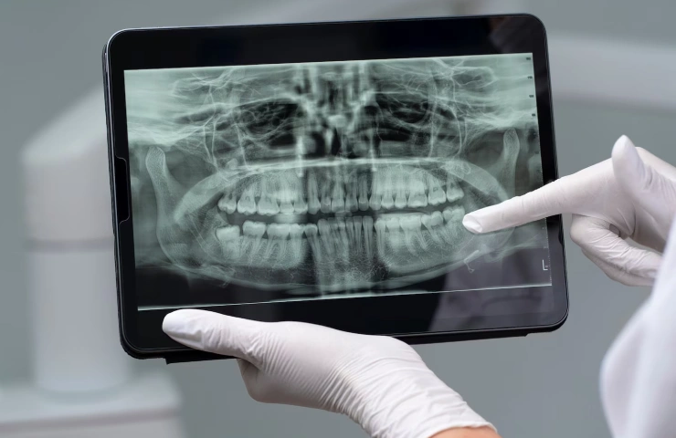
Maxillofacial surgery entails carefully planned interventions on the jaw, face, and related structures. Success in these procedures depends heavily on accurate imaging for diagnosis, surgical preparation, and post-operative review. Computed Tomography (CT) scans have emerged as essential tools due to their ability to generate detailed three-dimensional imagery. Nevertheless, the risks associated with radiation exposure have led to the development of alternative imaging options and improved strategies for optimizing scan protocols. This blog highlights the significance of CT scans, reviews safer imaging alternatives, and offers guidelines for obtaining CT scans with optimal slice thickness.
Why CT Scans Are Essential in Maxillofacial Surgery
CT scans are vital for maxillofacial surgery, offering unparalleled benefits:
- Fine-Detail Imaging
Maxillofacial anatomy is intricate, requiring imaging capable of capturing fine details. CT scans provide high-resolution cross-sectional views, enabling clinicians to assess the relationships between bone structures, air spaces, and soft tissues. These images are indispensable for identifying fractures, deformities, and other conditions.
- Multi-Dimensional Visualization
Unlike conventional X-rays, CT scans deliver three-dimensional reconstructions, allowing surgeons to examine anatomical structures from various perspectives. This capability improves surgical planning and reduces risks during procedures.
- Precision in Surgical Planning
Pre-operative planning often necessitates thorough evaluations of trauma, congenital anomalies, or tumors. CT scans enable surgeons to simulate interventions, foresee challenges, and craft detailed surgical plans tailored to each patient.
- Effective Post-Surgical Assessment
Post-operative imaging plays a critical role in evaluating recovery, detecting complications, and monitoring the success of procedures. CT scans allow surgeons to confirm implant placements and check healing progress with accuracy.
- Implant Placement Guidance
CT scans provide valuable insights into bone density and alignment during implant procedures. Precise imaging prevents implant failures while ensuring stable and functional results for patients.
- Complex Case Management
For challenging cases, such as extensive trauma or facial asymmetry, CT scans offer detailed visualizations necessary for planning and executing complex surgeries. Combined with advanced design tools, they help surgeons achieve optimal outcomes.
Safer Alternatives to Reduce Radiation Exposure
Minimizing radiation exposure remains a priority, and several imaging alternatives address this concern:
- Cone Beam Computed Tomography (CBCT)
CBCT has gained popularity for its ability to produce detailed images with much lower radiation doses compared to traditional CT scans. It is highly effective for localized imaging needs, such as dental assessments.
- Magnetic Resonance Imaging (MRI)
Using magnetic fields instead of radiation, MRI is ideal for visualizing soft tissues. While it isn't suitable for detailed bone imaging, it complements CT scans in cases involving vascular or nerve assessments.
- Ultrasound Imaging
Ultrasound is a safe and cost-effective option for examining superficial tissue structures. Though limited in visualizing deeper anatomy, it can be useful in specific applications like guided biopsies.
- Advanced Digital X-rays
Digital X-rays equipped with collimation features reduce unnecessary exposure by focusing the beam precisely on the target area. They are efficient for quick localized evaluations.
- Dose Optimization Techniques
Innovative imaging protocols, such as iterative reconstruction and adjustments to tube current, allow for reduced radiation exposure while maintaining image quality.
- Hybrid Imaging Modalities
Technologies like PET-CT or SPECT-CT combine functional and anatomical imaging, tailoring radiation doses for complex diagnostic scenarios.
Guidelines for Procuring CT Scans with Slice Thickness
Optimal slice thickness is a critical factor in ensuring high-quality imaging. Follow these guidelines:
- Define Clinical Objectives
The purpose of the scan dictates the appropriate slice thickness. Thin slices (0.5–1.0 mm) are ideal for detailed evaluations, while thicker slices (2–5 mm) suffice for broader assessments.
- Balance Resolution and Safety
Thinner slices improve resolution but may require higher radiation doses. Employing dose optimization techniques can maintain image quality while minimizing risks.
- Equip with Adaptive Protocols
Modern CT machines capable of adjusting slice thickness based on clinical needs are essential for efficient imaging.
- Position Patients Properly
Accurate positioning and immobilization during scans help reduce motion artifacts, preserving the integrity of images.
- Collaborate with Radiologists
Radiologists assist in selecting protocols that align imaging settings, slice thickness, and dose parameters with clinical objectives.
- Comply with Regulatory Standards
Adherence to established safety guidelines is mandatory to prevent excessive radiation exposure.
- Integrate Surgical Planning Tools
Ensure compatibility between CT systems and planning software for seamless integration of imaging data into surgical workflows.
Conclusion
CT scans are invaluable tools for maxillofacial surgery, enabling precise diagnosis and surgical planning for complex cases. Safer alternatives such as CBCT, MRI, and ultrasound offer viable solutions to minimize radiation risks, while advanced optimization techniques enhance the safety of CT imaging. By adhering to guidelines for slice thickness and leveraging technological advancements, healthcare professionals can achieve a balance between diagnostic excellence and patient safety. As imaging technologies continue to evolve, the future of maxillofacial surgery promises greater precision and innovation in patient care.
Healthcare providers must remain informed about imaging advancements and prioritize patient safety in every stage of treatment planning. A thoughtful approach to imaging ensures better clinical outcomes and reinforces trust in medical care.

Dr. Shilpi Bhadani
MBBS, MS - General Surgery, MCh - Plastic & Reconstructive Surgery,
DAFPRS Fellowship in Aesthetic Surgery
Recent Blog Posts
Non-Surgical Gynecomastia Treatment: Does It Work?
June 25, 2026
SB Aesthetics is one of the renowned medical centers in Gurgaon offering world-class and most advanced plastic surgeries procedures under the guidance of Dr. Shilpi Bhadani.
Request An Appointment
Last reviewed: June 2026
Medically reviewed by Dr. Shilpi Bhadani.
Copyright © 2021-2026, SB Aesthetics. All Rights Reserved. Powered by DigiLantern